The Impossible Eye
Seeing is Believing: The Design of the Human Eye
If one of your friends asked you, “How do you know God exists?,” what would you say? There are many different ways to prove God’s existence, because God has given us so much evidence. Sometimes we find that evidence in things we see in the Universe, for example, the Sun. The Sun is like a giant nuclear engine. It gives off more energy in a single second than mankind has produced since the Creation. It converts 8 million tons of matter into energy every single second, and has an interior temperature of more than 20 million degrees Celsius (see Lawton, 1981). Sometimes we find evidence in the animal kingdom. Take the golden orb spider for instance. Pound for pound, the dragline silk of this spider is five times stronger than steel, and is twice as strong as the material that currently makes upSWAT teams’ bulletproof vests. In fact, due to its amazing strength and elasticity, it has been said that you could trap a jumbo jet with spider silk that is the thickness of a pencil.
One of the best examples of design within the human body is the eye. Even Charles Darwin struggled with the problem of how to explain how such a complex organ as the eye could have “evolved” through naturalistic processes. In The Origin of Species he wrote:
To suppose that the eye with all its inimitable contrivances for adjusting the focus to different distances, for admitting different amounts of light, and for the correction of spherical and chromatic aberration, could have been formed by natural selection, seems, I freely confess, absurd in the highest sense (1859, p. 170, emp. added).
But even though Darwin acknowledged that the eye could not have evolved, he went on to argue that it had, in fact, been produced by natural selection through an evolutionary process. It seems almost as though Darwin could not seem to make up his mind on the matter. But he is not the only one who has struggled to explain, from a naturalistic viewpoint, the intricacy of the eye. Evolutionist Robert Jastrow once wrote:
The eye is a marvelous instrument, resembling a telescope of the highest quality, with a lens, an adjustable focus, a variable diaphragm for controlling the amount of light, and optical corrections for spherical and chromatic aberration. The eye appears to have been designed; no designer of telescopes could have done better. How could this marvelous instrument have evolved by chance, through a succession of random events? (1981, pp. 96-97, emp. added).
How indeed? Though Dr. Jastrow argued that “the fact of evolution is not in doubt,” he confessed that “…there seems to be no direct proof that evolution can work these miracles.… It is hard to accept the evolution of the eye as a product of chance” (1981, pp. 101,97,98, emp. added). Considering the extreme complexity of the eye, it is easy to understand why Jastrow would make such a comment. In his book, Does God Believe in Atheists?, John Blanchard described just how complex the eye really is.
The human eye is a truly amazing phenomenon. Although accounting for just one fourth-thousandth of an adult’s weight, it is the medium which processes some 80% of the information received by its owner from the outside world. The tiny retina contains about 130 million rod-shaped cells, which detect light intensity and transmit impulses to the visual cortex of the brain by means of some one million nerve fibres, while nearly six million cone-shaped cells do the same job, but respond specifically to colour variation. The eyes can handle 500,00 messages simultaneously, and are kept clear by ducts producing just the right amount of fluid with which the lids clean both eyes simultaneously in one five-thousandth of a second (2000, p. 313).
Statements like this proves that the eye was so well designed, and so complicated, that it could not have happened by accident, as evolution teaches.
THE EYE’S DESIGN
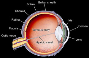
The anatomy of the eye was first examined and recorded at Alexandria, Egypt, in the first century A.D. An anatomist, Rufus of Ephesus, described the main parts of the eye, which included the dome-like cornea at the front, the colored iris, the lens, and the vitreous humor (which gives the eye its shiny look). Today, thanks to microscopes, we now know that these, along with many other parts of the eye, work in harmony to produce the gift of sight.
The outer white layer of the eye is called the sclera, more commonly known as the “white of the eye.” This layer is an extremely durable, fibrous tissue that extends from the cornea (the clear front section of the eye) to the optic nerve (at the back of the eye). Six tiny muscles (known as the extraocular muscles, or EOMs) connect to the sclera around the eye and control the eye’s movements. Four of the muscles (known as the rectus muscles) control the horizontal and vertical movement, while two (the oblique muscles) control the rotation. All six muscles work together so that the eye moves smoothly.
The inside of the eye can be divided functionally into two distinct parts. The first is the physical “dioptric” mechanism (from the Greek word dioptra, meaning something through which one looks), which handles incoming light. This includes the cornea, iris, and lens. The cornea is the transparent, dome-shaped window (about eleven millimeters in diameter) that covers the front of the eye. Its most important function is to protect the delicate components of the eye against damage by foreign bodies. Thus, the cornea acts like a watch face, in that it lets us look through the “window” of our eye while protecting the internal components from debris and harmful chemicals. The cornea also takes care of most of the refraction (the ability of the eye to change the direction of light in order to focus it on the retina) and works with the lens to help focus items seen at varying distances as it changes its curvature. The iris and the pupil work together to let in just the right amount of light. There are two opposing sets of muscles that regulate the size of the aperture (the opening, or the pupil) according to the brightness or dimness of the incoming light. If the light is bright, the iris constricts, allowing little light to pass; but if it is dark, the iris dilates or expands, allowing more light to pass through. The light (or image) then moves through a lens that has the ability to adjust its shape to help it clarify the image by changing the focal length of the lens between 40.4 and 69.9 millimeters where it is then focused (in an inverted form) on to the retina.
Between the lens and the retina is a transparent substance (the vitreous fluid) that fills the center of the eye. This substance is important because it not only gives the eye its spherical shape, but also provides nutrition for the retinal vessels inside the eye. In children, the vitreous feels like a gel, but as we age, it gradually thins and becomes more of a liquid.
The second is the receptor area of the retina where the light triggers processes in the nerve cells. The retina plays a key role in visual perception. In his book, The Wonder of Man, Werner Gitt explains how the retina is a masterpiece of engineering design.
One single square millimetre of the retina contains approximately 400,000 optical sensors. To get some idea of such a large number, imagine a sphere, on the surface of which circles are drawn, the size of tennis balls. These circles are separated from each other by the same distance as their diameter. In order to accommodate 400,000 such circles, the sphere must have a diameter of 52 metres… (1999, p. 15).
Alan L. Gillen also praised the design of the retina in his book, Body by Design.
The most amazing component of the eye is the “film,” which is the retina. This light-sensitive layer at the back of the eyeball is thinner than a sheet of plastic wrap and is more sensitive to light than any man-made film. The best camera film can handle a ratio of 1000-to-1 photons in terms of light intensity. By comparison, human retinal cells can handle a ratio of 10 billion-to-1 over the dynamic range of light wavelengths of 380 to 750 nanometers. The human eye can sense as little as a single photon of light in the dark! In bright daylight, the retina can bleach out, turning its “volume control” way down so as not to overload. The light-sensitive cells of the retina are like an extremely complex high-gain amplifier that is able to magnify sounds more than one million times (2001, pp. 97-98, emp. added).
Without a doubt, this thin (only 0.2 mm) layer of nerve tissue is a marvel of engineering. It contains photoreceptor (light-sensitive) cells and four types of nerve cells, as well as structural cells and epithelial pigment cells. The two kinds of photoreceptor cells are referred to as rods and cones because of their shape. Each eye has about 130 million rods and 7 million cones. The rods are very sensitive to light (whether it is bright or dim), and allow the eye to see in black and white. Cones, on the other hand, are not as sensitive as rods, and function only optimally in daylight. There are three different types of cones—red light, green light, and blue light—each of which is sensitive to its respective color of light, and which allow the eye to see in full color. The rods and cones convert the different lights into chemical signals, which then travel along the optic nerve to the brain.
Not only are the images produced by the dioptric mechanism miniaturized and upside-down, but it turns out that they also are left-right inverted. The optic nerves from both eyes split up and cross each other in such a way that the left halves of the images of both eyes are received by the right hemisphere of the brain, while the right halves are received by the left. Each half of the observer’s brain receives information from only one half of the image. As Gitt went on to explain, “Note that, although the brain processes the different parts of the image in various remote locations, the two halves of the field of vision are seamlessly reunited, without any trace of a joint—amazing! This process is still far from being fully understood” (p. 17). It is hard to believe that this inverted system of sight could have been produced through evolution.
Since the eyes are one of the most important organs in the body, they must be taken care of constantly. And God designed just such a built-in cleaning system, consisting of the eyelashes, eyelids, and lacrimal glands. The lacrimal glands produce a steady flow of tears that flush away dust and other foreign materials. The tears also contain a potent anti-microbial agent known as lysozyme that destroys bacteria, viruses, etc. The eyelids and eyelashes work together to keep dirt and other debris from entering the eye. The eyelids act like windshield wipers, blinking 3-6 times a minute to moisten and clean the eye.
For many years, scientists have compared the eye to the modern manmade camera (see Miller, 1960, p. 315; Nourse, 1964, p. 154; Gardener, 1994, p. 105). True, the eye and camera do have many things in common, if the function of the camera demands that it was “made,” does it not stand to reason that the more complex human camera, the eye, also must have had a Maker? Alan Gillen explained it best when he wrote: “No human camera, artificial device, nor computer-enhanced light-sensitive device can match the contrivance of the human eye. Only a master engineer with superior intelligence could manufacture a series of interdependent light sensitive parts and reactions”


